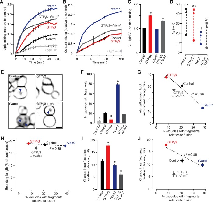
FIGURE 2:
GTPγS and rVam7 have opposing effects on the time interval between stalk and pore formation, fragment formation, and surface area during vacuole fusion. Lipid mixing (A) or content mixing (B) was recorded during in vitro homotypic vacuole fusion reactions in the absence (control) or presence of 100 nM rVam7, 0.2 mM GTPγS, or both (n ≥ 6). As negative control, 3.2 µM Gyp1-46, a Rab-GTPase inhibitor, was added to reactions to prevent fusion. (C) Maximum lipid mixing and content mixing values observed as ratios for each condition. (D) Times at half-maximal lipid mixing (circles) and content mixing (squares); time intervals between events were calculated (numbers) and are shown (arrow). (E) Images of FM4-64 stained vacuoles obtained 60 min after fusion was initiated. Arrowheads indicate intralumenal fragments. Scale bar, 2 µm. Using micrographs of fusion reactions, we calculated the percentage of vacuoles with intralumenal membrane fragments (F) and compared these values to time intervals between stalk and pore formation (G), boundary length (H), or the change in surface area relative to number of fusion events (as assessed by content mixing; I, J) for each condition (n = 324–802). Asterisks indicate data points significantly different from control (p < 0.05).

