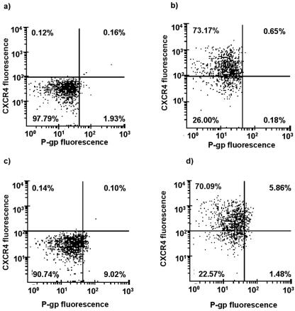
FIG. 2.
Representative scatter plots obtained from dual-color flow cytometry of PBMC stained with (a) isotypically matched negative-control antibodies and with (b) CXCR4-specific, (c) P-gp-specific, and (d) P-gp- and CXCR4-specific antibodies. Values in each quartile indicate number of events within the region.

