FIG.3.
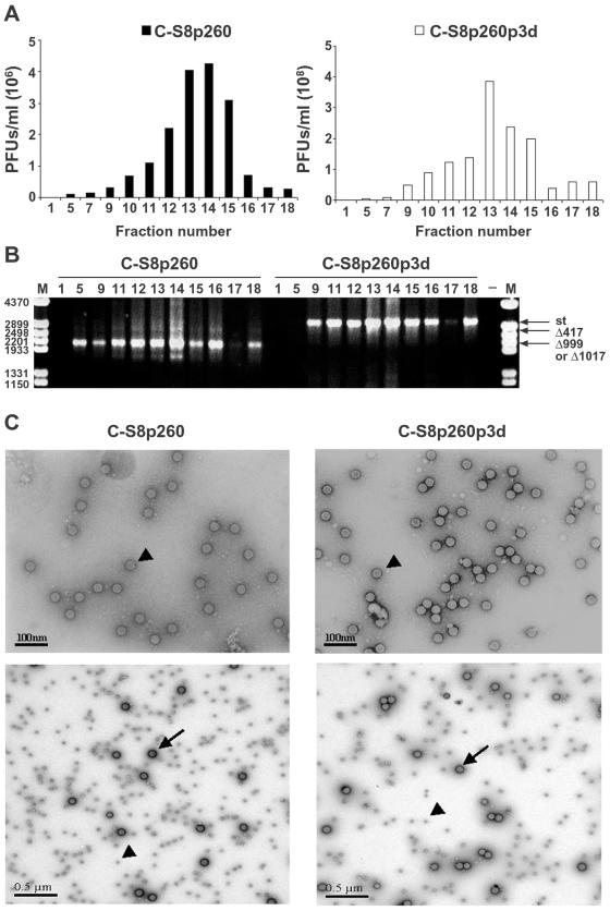
Analysis of encapsidation of viral genomic RNAs from FMDV C-S8p260 and C-S8p260p3d. (A) Sedimentation analysis of C-S8p260 and C-S8p260p3d. Sucrose density gradient infectivity profile of FMDV preparations C-S8p260 and C-S8p260p3d. Viral particles were purified as described in Materials and Methods. Infectivity was measured by titration of each gradient fraction. Fraction 1 corresponds to the top and fraction 18 to the bottom of the gradients. Each value represents the mean of triplicate assays and standard deviations (data not shown) never exceeded 15%. (B) Agarose gel electrophoresis of the products of RT-PCR amplifications of FMDV RNAs extracted from fractions of the sucrose gradient sedimentation of C-S8p260 and C-S8p260p3d. Gradient fractions from which the RNAs were analyzed correspond to those shown in panel A, and the fraction number is indicated above each lane. Amplification 21 (Fig. 1A) was used. Input RNA was diluted 100 times to make it limiting for the amplification reaction so that the amount of PCR product obtained was proportional to the input template RNA. Amplification products corresponding to the standard (st) and to the RNAs with deletions (Δ417 and Δ999 or Δ1017) are shown. Lane M, molecular size markers (HindIII-digested φ29DNA; the corresponding sizes [base pairs] are indicated on the left); lane −, negative control without RNA. (C) Electron microscopy photographs of viral populations C-S8p260 and C-S8p260p3d. Purified virus from fraction 14 of the sucrose gradients (Fig. 3A) was analyzed by electron microscopy. Negatively stained viral particles (examples indicated by arrowheads) at a magnification of ×25,000 (top frames). The measured size of the viral particles in both populations was 30 ± 0.1 nm (average of 30 measurements). Photographs of viral particles (examples indicated by arrowheads) mixed with 91-nm latex beads (examples indicated by arrows) of known size and concentration at a magnification of ×8,000 (bottom frames). C-S8p260 was not diluted; C-S8p260p3d was diluted 5 times; latex beads were diluted 20 times in both cases. Methods are detailed in Materials and Methods.

