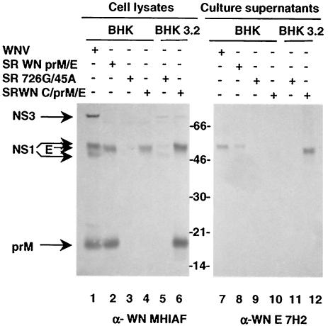
FIG. 2.
Analysis of intracellular and secreted WNV antigens. BHK or BHK3.2 cells were infected with WNV (lanes 1 and 7) or transfected with RNAs transcribed from the indicated constructs. Cells and supernatants were harvested 30 h after electroporation and analyzed by immunoblot. (Left) Antigen expression in cell lysates detected with polyclonal MHIAF; (right) antigens secreted into the culture supernatant, detected with a monoclonal E-specific antibody. Note that E is secreted only from WNV-infected cells, BHK cells transfected with SR WN prM/E RNA, and BHK3.2 cells transfected with SR WN C/prM/E but not parental BHK cells transfected with SR WN C/prM/E.

