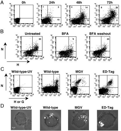
FIG. 1.
MV N is expressed at the cell membranes of infected PBLs. (A) Human PBLs were infected with MV strain Edmonston (MOI, 0.5), and the cell surface expression of N and H was analyzed by immunocytometry at different times after infection. The percentage of positive cells is given in the corner of each quadrant. (B) PBLs were infected with MV Edmonston; 48 h later, they were incubated with BFA (1 μg/ml) for 4 h; afterwards they were either stained or washed thoroughly, incubated for an additional 2 h at 37°C, and stained. (C) PBLs were infected with wild-type MV strain G954 (MOI, 0.1), previously inactivated or not by UV irradiation, or with the H- and F-deficient recombinant MV strain (MGV) or its molecularly cloned control (EDtag) (MOI, 0.5). N and H expression was analyzed 48 h postinfection. An isotype control for anti-N staining was used in all experiments; less than 0.5% of cells were positive with this control. (D) Immunofluorescence staining of N on nonpermeabilized human PBLs infected with wild-type MV, merged with transmitted light image.

