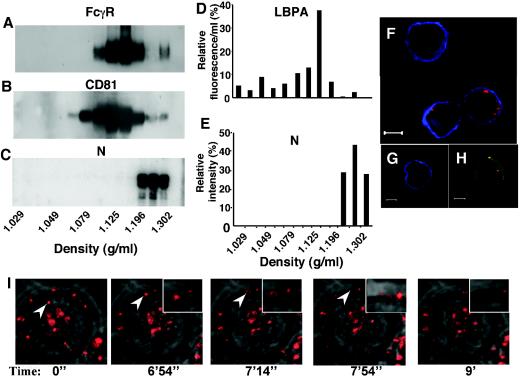
FIG. 4.
MV N is secreted and binds to neighboring cells. P815-N supernatants were subjected to CsCl gradients. After centrifugation, fractions were collected and analyzed by Western blotting for (A) FcγR, (B) CD81, and (C) N. (D) LBPA content in different fractions was determined by ELISA. (E) N content in different fractions was evaluated by phosphorimager. (F-H) P815-M cells, cultured 48 h with P815-N cells in transwell cultures (F and H) or alone (G), were analyzed for expression of FcγR, MV N, and MV M protein by confocal microscopy. N and FcγR expression is visualized by red and green, respectively. (F, G) M expression was analyzed after cell permeabilization and is visualized in blue. (H) Yellow in merged panels shows colocalization of N and FcγR. (I) Dynamics of cellular transit of N. Images are extracted from time-lapse video analysis of P815-N cells stained by an anti-N MAb, merged with a transmitted light image. Arrowheads follow a representative N-labeled spot up to its release from the cell surface and dispersion in the extracellular milieu.

