FIG. 5.
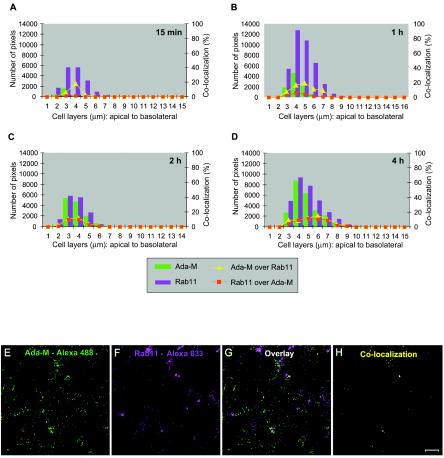
HIV-1 colocalizes weakly with recycling endosomes. Polarized JAR cells were colabeled with purified human anti-HIV-1 followed by an Alexa 488-conjugated goat anti-human antibody and rabbit anti-Rab11 followed by Alexa Fluor 633-conjugated goat anti-rabbit IgG. (A to D) Bars indicate the numbers of HIV-1-specific (green) and Rab11-specific (magenta) pixels over the entire depth of the cells 15 min, 1 h, 2 h, or 4 h following exposure to HIV-1. Curves represent the percentage of colocalization of Ada-M versus Rab11 (yellow triangles) or Rab11 versus Ada-M (red squares) over the entire depth of the cells. (E to H) Digital images of the 4-μm cell layer following a 1-h exposure to HIV-1. (E) Ada-M is shown in green. (F) Rab11 is depicted in magenta. (G) Overlay of signals specific for HIV-1 and Rab11. (H) Binary image showing the regions of colocalization between HIV-1 and Rab11 on this cell layer (yellow dots). Bar, 10 μm.

