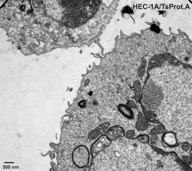
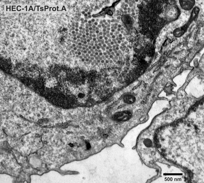
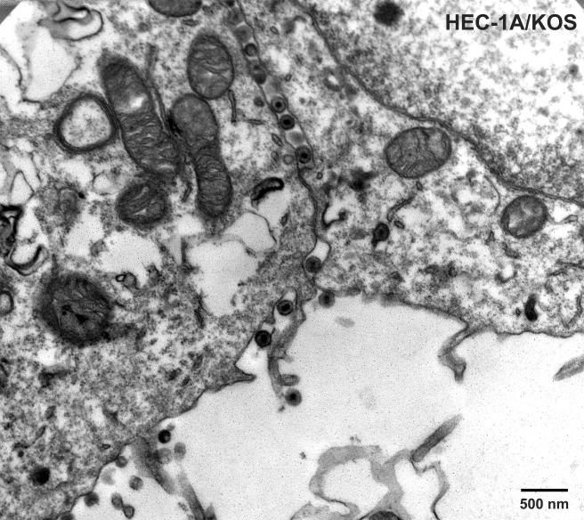
FIG. 1 to 3.
Electron microscopic examination of HEC-1A cells infected with TsProt.A and wild-type HSV strain KOS. HEC-1A cells were infected with TsProt.A (Fig. 1 and 2) or KOS (Fig. 3) at 39°C for 18 to 19 h. The cells were washed once with PBS+/+, fixed with glutaraldehyde for 30 min while still affixed to plastic dishes, washed twice with sodium cacodylate buffer, scraped, and collected by centrifugation at 1,000 × g for 5 min. The cell pellet was postfixed and embedded in epoxy, and ultrathin sections were stained with uranyl acetate and lead citrate and then viewed in an electron microscope. Bar, 500 nm.


