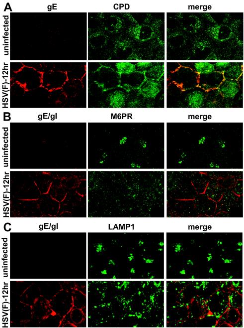
FIG. 10.
Subcellular distribution of cellular proteins CPD, M6PR, and LAMP1 in HSV-infected HEC-1A cells. HEC-1A cells were infected with wild-type HSV-1 strain F then fixed and processed as described Fig. 4. The cells were then stained with the following antibodies: rabbit anti-CPD and anti-gE mouse MAb 3114, followed by Alexa 594-labeled goat anti-rabbit IgG and Alexa 488-labeled goat anti-mouse IgG (A); mouse anti-M6PR and rabbit anti-gE/gI polyclonal antibodies, followed by Alexa 488-labeled goat anti-mouse IgG and Alexa 594-labeled goat anti-rabbit IgG (B); or mouse anti-LAMP1 and rabbit anti-gE/gI, followed by Alexa 594-labeled goat anti-rabbit IgG and CY5 goat anti-mouse IgG (C). For clarity, in each case the gE or gE/gI is shown in red, and CPD, M6PR, or LAMP1 is shown in green. Thus, in panel A the red and green channels were switched, and in panel C the blue (CY5) channel was converted to green.

