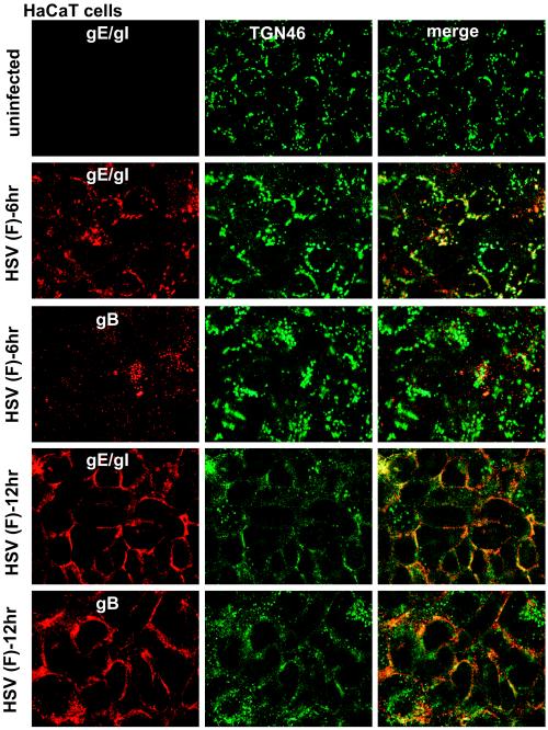
FIG. 12.
Redistribution of gE/gI, gB, and TGN46 to cell junction in HaCaT keratinocytes. HaCaT cells were infected with wild-type HSV (F) for 6 or 12 h, fixed, permeabilized, and stained with rabbit anti-gE/gI (red) and sheep anti-TGN46 (green) antibodies, followed by Alexa 594-conjugated goat anti-rabbit IgG and Alexa 488-conjugated donkey anti-sheep IgG. Alternatively, cells were stained with mouse anti-gB MAb 15βB2 and I-59 (red) and sheep anti-TGN46 (green), followed by secondary Alexa 594-conjugated goat anti-mouse IgG and Alexa 488-conjugated donkey anti-sheep IgG.

