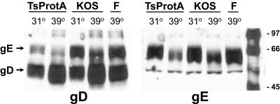
FIG. 6.
Cell surface expression of gE and gD in cells infected with TsProt.A. HEC-1A cells were infected with wild-type HSV strains KOS or F or with TsProt.A for 18 h at either 31 or 39°C. The cells were washed with cold PBS, cell junctions were disrupted by incubation with 2 mM EGTA for 25 min at 4°C, and then cells incubated with NHS-SS-biotin to biotinylate cell surface proteins. Unreacted biotinylation reagent was quenched, cells were lysed, and gD (left panel) or gE (right panel) was immunoprecipitated with rabbit anti-gD polyclonal sera or mouse anti-gE MAb 3114, respectively. The immunoprecipitated proteins were subjected to electrophoresis on sodium dodecyl sulfate-polyacrylamide gels, and then the proteins were transferred to membranes and incubated with streptavidin-conjugated peroxidase and chemiluminescence reagent. Images were captured on X-ray film. The positions of molecular weight markers and of gD and gE are indicated.

