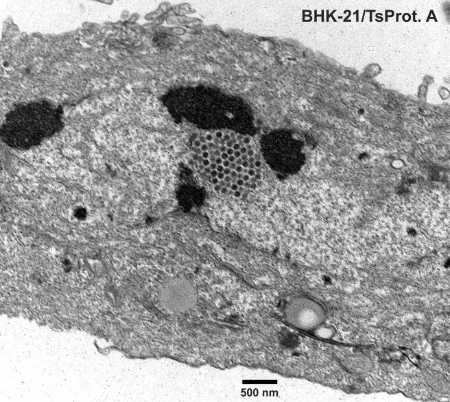
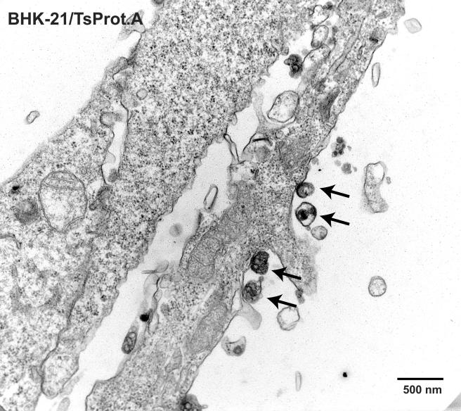
FIG. 7 and 8.
Nuclear capsids and cell surface L particles in TsProt.A-infected BHK-21 cells. BHK-21 cells were infected with TsProt.A at 39°C for 18 to 19 h. The cells were washed, fixed, and processed for electron microscopy as described in Fig. 1 to 3. Bar, 500 nm. Arrows in Fig. 8 indicate cell surface L particles.


