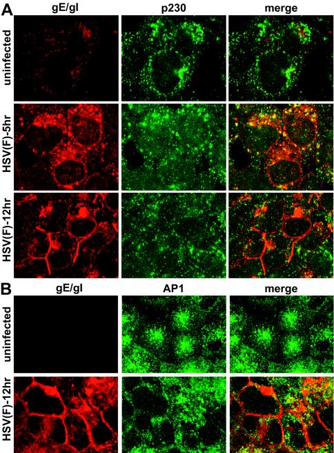
FIG. 9.
Subcellular distribution of TGN-resident cellular proteins p230 and AP-1 in HSV-infected epithelial cells. HEC-1A cells were left uninfected or infected with HSV-1 (strain F) for 6 or 12 h; the cells were then fixed with paraformaldehyde, permeabilized, blocked, and stained with mouse anti-p230 and simultaneously with rabbit anti-gE/gI polyclonal antibodies (A) or mouse anti-AP-1 and simultaneously with rabbit anti-gE/gI antibodies (B). The cells were washed and incubated with Alexa 488-conjugated goat anti-mouse IgG and Alexa 594-conjugated goat anti-rabbit IgG. The images show gE/gI as red (Alexa 594) and AP-1 or p230 as green (Alexa 488).

