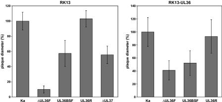
FIG. 3.
Plaque sizes of PrV-ΔUL36F, PrV-UL36BSF, PrV-UL36R, PrV-ΔUL37, and PrV-Ka. Virus plaques were visualized 48 h after infection of RK13 or RK13-UL36 cells by indirect immunofluorescence with a gC-specific antibody. For each virus, the average diameters of 30 plaques were determined and calculated as percentages of those of the plaques induced by PrV-Ka. Standard deviations are also shown.

