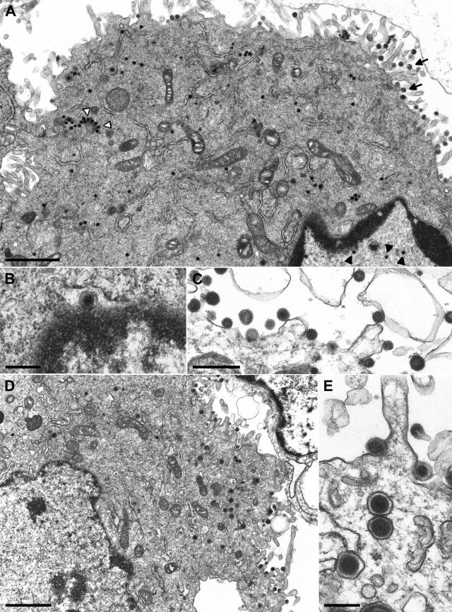
FIG. 5.
Electron microscopy of cells infected with phenotypically complemented PrV-ΔUL36F. RK13 (A to C) and RK13-UL36 cells (D, E) were analyzed 12 h after infection at a multiplicity of infection of 1. In RK13 cells, DNA-filled capsids were detected in the nucleus (A, black arrowheads) and in the cytoplasm (A, white arrowheads), and primary enveloped virus particles were found in the perinuclear space (B). However, only capsidless particles were efficiently released from the cells (A, arrows; C). In RK13-UL36 cells, enveloped virions were detectable in cytoplasmic vesicles and in the extracellular space (D, E). Bars: 1.5 μm (A, D), 500 nm (C), or 250 nm (B, E).

