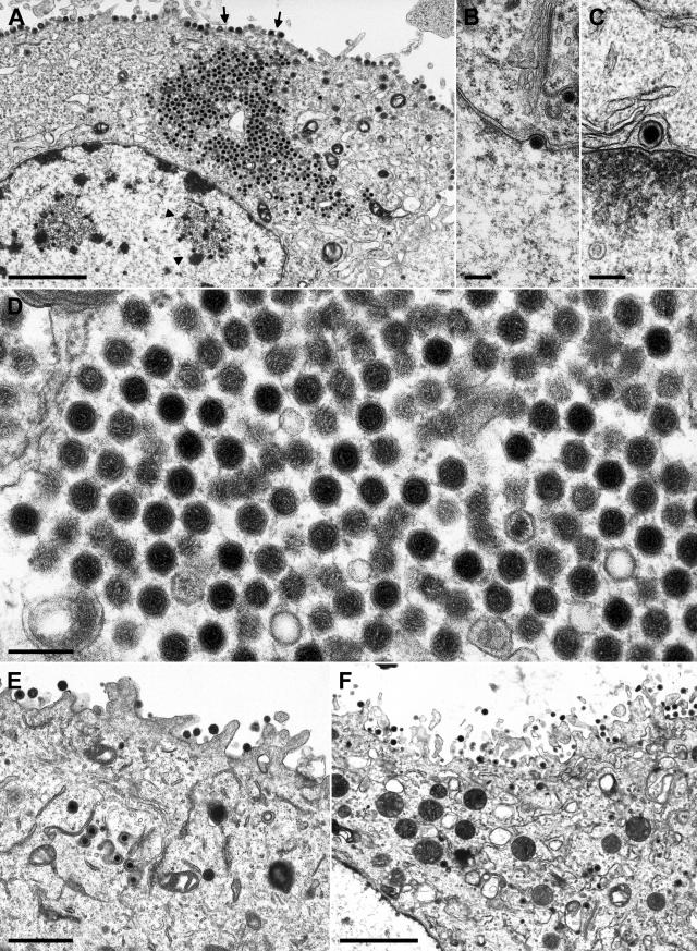
FIG. 6.
Electron microscopy of cells infected with PrV-UL36BSF. RK13 (A to D) and RK13-UL36 (E, F) cells were analyzed 12 h after infection. An overview (A) shows single intranuclear (arrowheads) and aggregated cytoplasmic nucleocapsids as well as capsidless extracellular particles (arrows). Enlargements depict primary envelopment at the nuclear membrane (B, C) and ordered clusters of unenveloped nucleocapsids in the cytoplasm (D). In RK13-UL36 cells, secondary envelopment was observed in the cytoplasm (E) and virions were released from the cells (E, F). Bars: 2.0 μm (A, F), 1.0 μm (E), or 200 nm (B, D).

