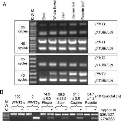
Figure 5.
Qualitative multiplex RT-PCR analysis of RNA transcripts extracted from various tissues and CAPS of PIMT2 splicing variants. A, The RT-PCR products at 25 and 40 reaction cycles for target cDNAs (PIMT1 and PIMT2) and the β-TUBULIN cDNA were separated on 1.5% (w/v) agarose gels. B, Amplicons of PIMT2 were not (−) or were (+) digested with Hpy188 III and PIMT2ψ and PIMT2ω splicing variant amounts visualized by ethidium bromide staining after electrophoresis through a 1.5% (w/v) agarose gel. The specificity of the CAPS analysis was demonstrated in the first two lanes using respective plasmid DNA templates under identical PCR and digestion conditions. The average ± se of multiple estimates of PIMT2ω abundance relative to total PIMT2 transcripts is indicated on the top of each lane.

