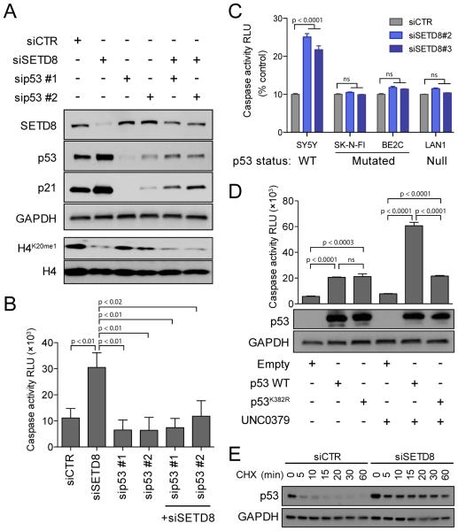
Figure 6. SETD8 inhibition-mediated cell death is p53 dependent.
(A) Immunoblot analysis showing p53 and/or SETD8 protein levels upon treatment with 2 different siRNAs targeting p53 alone or in combination with siSETD8 #3 for 48 hr in SY5Y cells.
(B) Caspase 3/7 activity calculated as RLU (Relative Luminescence Unit) upon treatment with 2 different siRNAs targeting p53 alone or in combination with siSETD8 #3 for 72 hr in SY5Y cells. Data represent mean ± SD of 2 independent experiments (p < 0.001).
(C) Caspase 3/7 activity calculated as RLU after SETD8 knockdown for 72 hr in two p53 mutated (SK-N-FI and BE2C) and in one p53 null (LAN1) NB cells compared with p53 WT SY5Y. Data represent mean ± SD of 2 independent experiments (p <0.001).
(D) Caspase 3/7 activity calculated as RLU with overexpression of p53 WT or p53K382R and treatment with UNC0379 8 μM for 24 hr in LAN1, p53 null NB cells. Data represent mean ± SD of 2 independent experiments (p < 0.001) (upper panel). Immunoblot analysis showing p53 levels under indicated conditions (lower panel).
(E) Immunoblot analysis for p53 with siCTR or siSETD8 in SY5Y cells. Cells were collected after treatment with 50 μg/ml CHX at the indicated time.
See also Figure S6.

