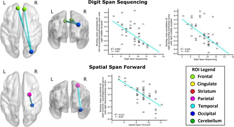
Fig. 1.

Relationship of working memory to resting-state brain connectivity. Digit Span Sequencing performance negatively regressed to resting-state functional connectivity of right middle occipital gyrus (bordering superior parietal lobule) to bilateral dorsomedial prefrontal cortex (top). Spatial Span Forward performance negatively regressed to functional connectivity of right fusiform/parahippocampal gyrus with right lateral premotor area (bottom). All depicted relationships survived FDR correction (q ≤ 0.05). Scatterplots indicate significant robust linear regressions between performance (abscissa) and functional connectivity (ordinate), with data points indicated by subject number (01-79). ROIs are color-coded by region for all figures: light green for prefrontal, yellow for cingulate, red for striatum, magenta for sensorimotor, cyan for temporal, blue for occipital, and dark green for cerebellum. Brain connectivity is depicted with BrainNet Viewer (Xia, Wang, & He, 2013).
