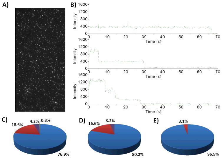
Figure 4.
Single-molecule photobleaching of variant CYP3A4 enzymes labelled with DyLight 549 maleimide (10). A) Representative example of a wide-field single molecule total internal reflection fluorescence (TIRF) microscope image obtained for fluorescently labelled CYP3A4 upon excitation at 532 nm. B) Representative intensity-time trajectories observed when the protein has one fluorophore/protein molecule (one step), two fluorophores/protein molecule (two steps) and three fluorophores/protein molecule (three steps). These trajectories were tallied for: CYP3A4 wild-type, n = 2212 (C); mutant 1, n = 1778 (D); and mutant 3, n = 703 (E). Pie colours used: blue = one step, red = two steps, purple = three steps, green = four steps.

