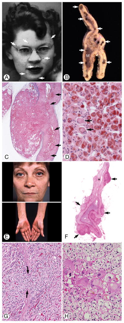
Figure 3.
Suspicious for Cushing Syndrome. A through D, Patient 28 (the index patient for Carney complex), E through H, Patient 27. A, The patient had spotty facial pigmentation (arrows). B, Cut surface of formalin-fixed adrenal gland obtained at autopsy showed brown micronodules (arrows) at junction of cortex and medulla (M). The normal shape of the gland was preserved. C, Sharply circumscribed mass of cortical cells mushroomed from the gland through a break in the capsule (stained blue with Masson trichrome technique). Multiple cortical micronodules were present (arrows) (original magnification ×40). D, A cortical micronodule was composed of round eosinophilic cells heavily stained with lipochrome. A few very large cells had little or no pigment (arrows) (hematoxylin-eosin, original magnification ×400). E, Spotty facial (upper) and forearm (lower) pigmentation and diffuse palmar pigmentation (lower). F, The cortex showed separation into zona fasciculata (vacuolated cells) and zona reticularis (eosinophilic cells). Multiple extra-adrenal mixed clear and eosinophilic ovoid cortical micronodules (arrows) indented the capsule. One intra-adrenal cortical micronodule was present (asterisk) (hematoxylin-eosin, original magnification ×1). G, Vacuolated cortical cells (right) extruded from the cortex though a break in the capsule (between arrows) to the exterior (hematoxylin-eosin, original magnification ×100). H, An aggregate of huge eosinophilic cortical cells surrounded by clusters of zona fasciculata-type vacuolated cells (hematoxylin-eosin, original magnification ×400).

