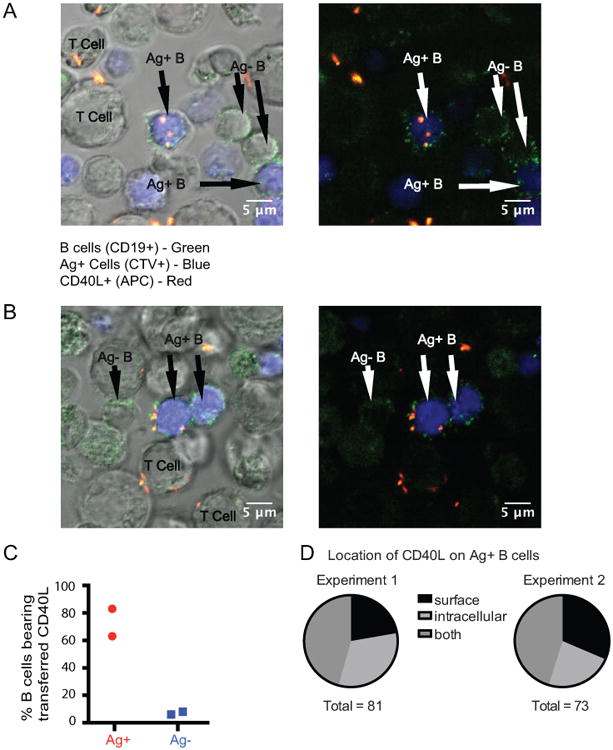
Figure 4. Transferred CD40L is found intracellularly and on the surface of antigen-pulsed B cells following overnight incubation with Th1 cells in the presence of anti-CD40L.

(A, B) Two representative images of Ag+ B cells (green (CD19+) and blue (CTV)) and bystander B cells (green only (CD19+)) together with T cells are shown with and without the differential interference contrast overlay following overnight incubation. The images in A and B show a frame through the middle of the cells from videos (Supporting Information Video 1 and Video 2) created from 2.5 μM z stacks. Intracellular CD40L (allophycocyanin-labeled anti-CD40L, shown in red) is detected in the Ag+ B cell in A and an example of surface CD40L is shown in B. (C) The graph shows the percentage Ag+ and Ag- B cells positive for the presence of CD40L in two individual experiments. (D) The pie charts show the relative numbers of B cells positive for surface, intracellular, or both surface and intracellular CD40L. There were 281 B cells analyzed in Experiment 1 (128 Ag+ and 153 Ag-) and 193 B cells in Experiment 2 (88 Ag+ and 105 Ag-).
