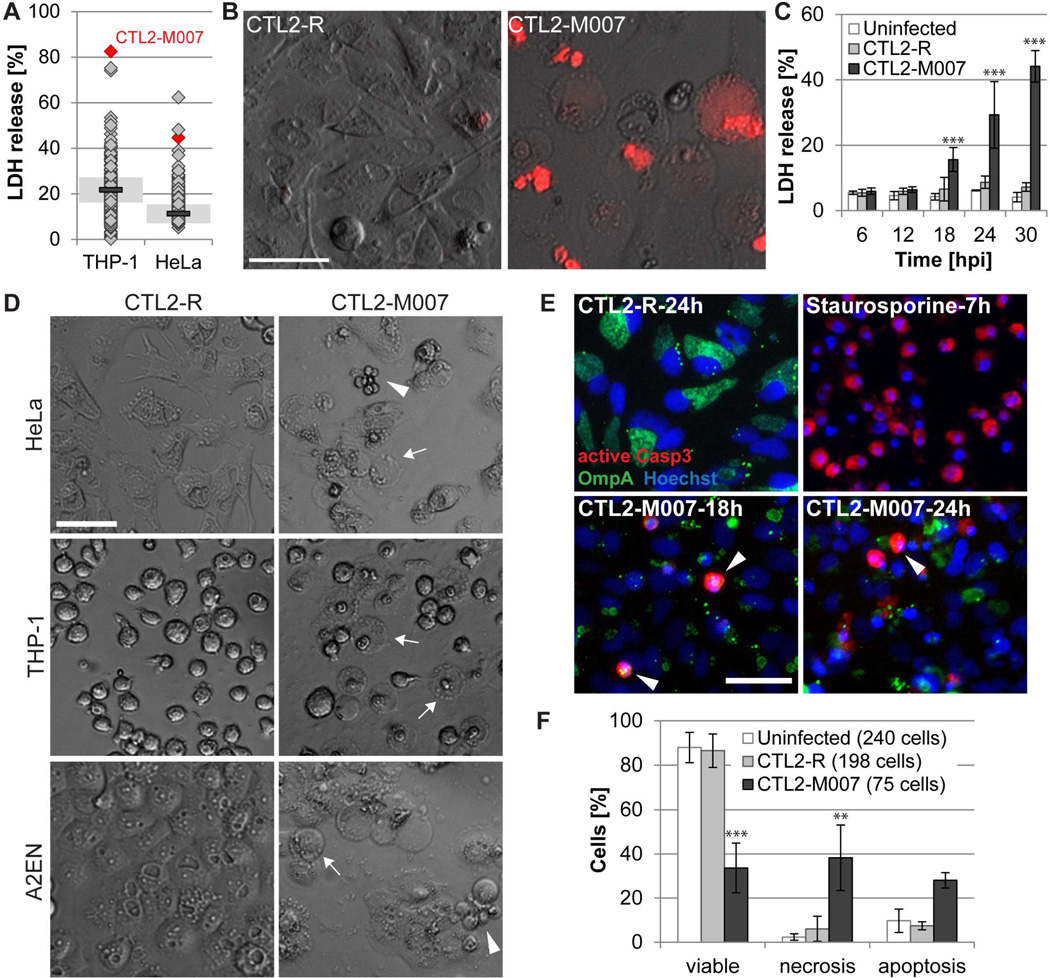
Figure 1. A genetic screen identifies a C. trachomatis strain that causes apoptotic and necrotic cell death.
(A) Cell lysis induced by Chlamydia mutants as assessed by the release of LDH into supernatants at 28 hpi (THP-1) or 24 hpi (HeLa) (mutants (diamonds, n=224); CTL2-R (bar, mean; shaded area, SD; n=2)).
(B) Loss of membrane integrity during infection of HeLa cells with CTL2-M007 (10 IFU/cell) visualized by enhanced permeability to propidium iodide (red) at 24 hpi (bar=50 µm).
(C) Time course of cell death induction by CTL2-M007 (10 IFU/cell) in HeLa cells (mean±SD, n=3, two-way ANOVA + Newman-Keuls).
(D) Induction of apoptosis (arrowheads) and necrosis (arrows) by CTL2-M007 in epithelial (HeLa, A2EN) and monocytic (THP-1) cells (10 IFU/cell, 21 hpi). Bar=50 µm.
(E) Immunofluorescence detection of apoptotic CTL2-M007-infected (10 IFU/cell) HeLa cells (Chlamydia OmpA, green; active caspase-3, red; Hoechst, blue). Arrowheads: apoptotic infected cells. Bar=50 µm.
(F) Live imaging based assessment of the frequency of apoptosis and necrosis in CTL2-M007-infected (10 IFU/cell) cells until 30 hpi. The category “uninfected” refers to cells in infected wells that contain no inclusions (mean±SD, n=2, two-way ANOVA + Newman-Keuls).
See also Fig. S1 and movies S1–3.

