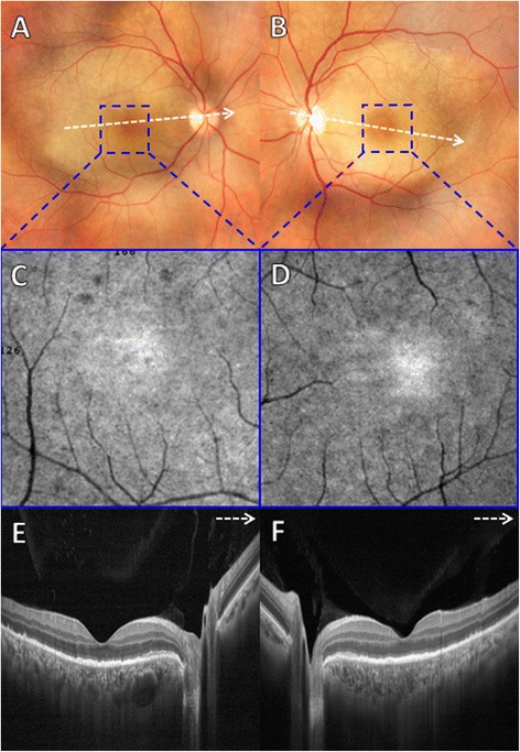
Fig. 2.

a, b Color fundus photograph of both eyes revealed a yellowish and placoid lesion within the posterior pole. c, d En-face OCT angiography 3 × 3 mm at the level of outer retina demonstrated multiple hyperreflective dots uniformly distributed within the foveal area of both eyes. e, f B-scan SD-OCT of both eyes revealed an intact ELM, disruption and loss of EZ, small nodular elevations on RPE and punctate hyperreflectivity in the choroid. g, h At 1-month follow-up, color fundus photograph showed disappearance of placoid lesion in both eyes. i, j At 1-month follow-up, en-face OCT angiography 3 × 3 mm at the level of outer retina showed partial disappearance of hyperreflective dots within the foveal area of both eyes. k, l At 1-month follow-up, B-scan SD-OCT of both eyes demonstrated remodeling of the EZ band and nearly total resolution of RPE nodular elevations and punctate hyperreflectivity in the choroid
