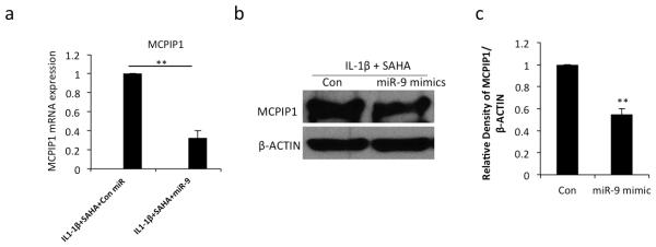
Figure 4. Overexpression of miR-9 suppress MCPIP1 expression in IL-1β stimulated and SAHA treated OA chondrocytes.
(a) Chondrocytes were transfected with control miR or miR-9 mimic (200nM) using Nucleofactor kit and were grown for 24 hrs and then treated with IL-1β (2ng/ml) and SAHA (1 μM). After 16 hrs of treatment total RNA was isolated and MCPIP1 expression was measured with β-actin as a normalizing control. (b) Chondrocytes were treated as above and subjected to Western blot analysis to detect the MCPIP1 and IL-6 proteins. β-actin was used as a loading control. Representative blot is shown. (c) Densitometric analysis of immunoreactive bands represented in (b) was performed. (*P <0.05; **P <0.005; paired student t-test).

