
Figure 1. Ion conductance and water permeability of Dx channels reconstituted in bilayers.
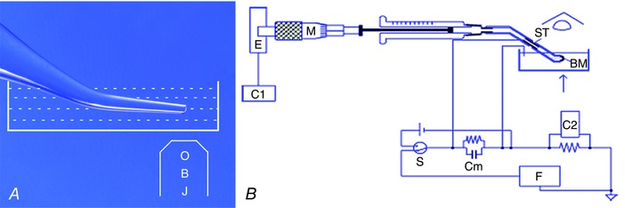
A, channel current recording: an aluminosilicate microelectrode (100 μm tip inner diameter) was bent on a microforge by 30 deg, silanized, filled with salt solution symmetrical with the bath, connected to a patch‐clamp amplifier headstage and immersed in a grounded salt solution in a 35 mm Petri dish on the stage of an inverted microscope. A bilayer membrane (BM) was formed at the tip of the microelectrode. OBJ, Objective lens B, water flow measurement: a BM was formed at the tip of a bent polytetrafluoroethylene (PTFE) tube filled with and immersed in KCl solution in a plastic Petri dish grounded with an Ag/AgCl wire. The back of the PTFE tube was connected through an Ag/AgCl tube (ST) to a Hamilton syringe whose plunger was driven by a micrometer (M), connected to an absolute encoder (E). The Ag/AgCl pair was used to set the voltage across the bilayer at zero. Water flow across the bilayer was first driven by a gradient of 500 mm urea. The membrane was kept at a fixed position of an eyepiece graticule of a microscope so that the rate at which the syringe plunger had to be moved to keep the membrane position constant was the rate of water flow across the BM. Data were acquired through computers C1 for water flow and C2 for membrane capacitance. Cm, membrane circuit analogue; F, triangular function generator; S, make‐before‐break switch to toggle between voltage source and function generator.