
Figure 3. Dx is less effective from the apical side (representative electrical traces).
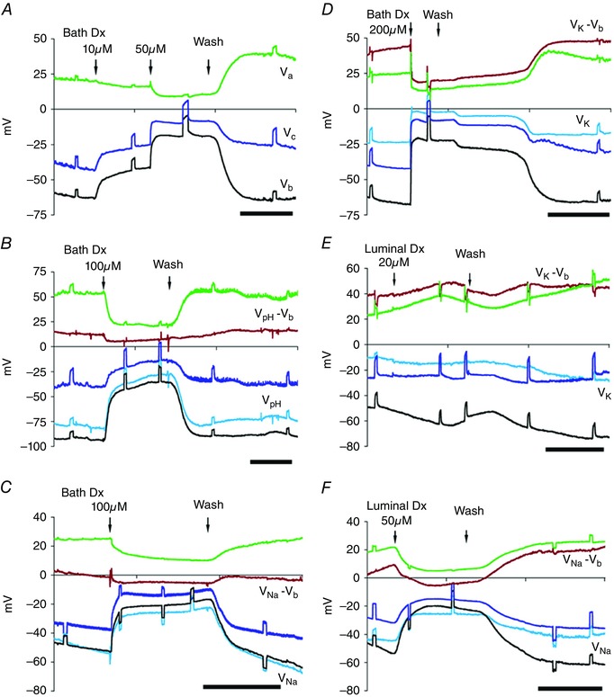
A–D, bath Dx: in Sch‐R, V c (blue trace), V b (black trace), V a (green trace) and intracellular ionic activities declined reversibly with graded doses of Dx. A, Dx 10 μm (threshold) depolarized V c and V b but not V a; higher doses (≥ 20 μm) depolarized V a (cf. A and D) reversibly for concentrations ≤ 100 μm; after wash both V a and V b hyperpolarized. B–D, depletion of intracellular H+ (B; see V pH – V b, brown trace), Na+ (C; see V Na – V b trace) and K+ (D; see V K – V b trace). E, luminal Dx of 20 μm has little effect; mild depolarization of the basal membrane V b and very little decrease in [K+]i (see V K – V b trace) was observed with no change in V c. F, luminal perfusion with 50 μm Dx depolarized V t or V c, V b and V a, and decreased [Na+]i (see V Na – V b trace). The short circuit current I sc was reduced without affecting R a/R b ratio (see Fig. 4 E and F). Scale bar, 5 min.