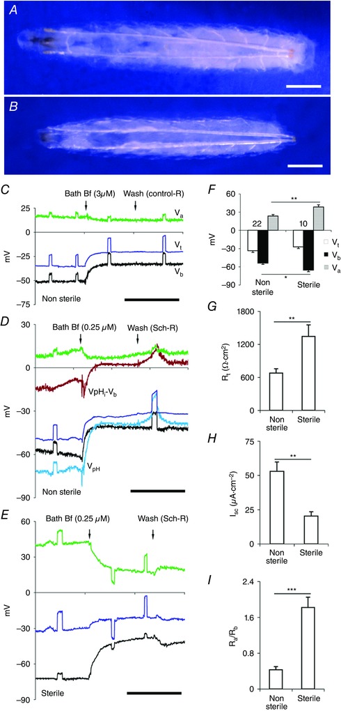
Figure 8. Sterile midguts have highly resistive apical membranes.

A, an actively feeding Drosophila CS larva (approximately 109 ± 3 h from egg laying) emerging into non‐sterile medium. B, Drosophila CS larvae of similar stage emerging into sterile medium show marginal reduction in size and attain this stage a day later than normal larvae (Non‐sterile, 109 ± 3 h vs. Gf, 129 ± 5 h; 25°C; n = 5 experiments). Scale bar, 0.25 mm. C–E, basal application of Bf in all panels. Scale bar, 5 min. C, in posterior midgut segment of wild‐type CS non‐sterile strains, with perfusion with control‐R (Table 1, solution a) V b depolarized much more than V a. D, perfusion with Sch‐R (Table 1, solution b), apart from the elevated depolarization effect of V c, V b but not V a, the intracellular H+ increased and pHi acidified (see V pH – V b, brown trace). E, sterile wild‐type CS posterior midgut with Bf; V a (green trace) and V b (black trace) depolarizations are comparable. F, apical potential Va and basal potential Vb of sterile larvae were hyperpolarized. G–I, sterile larvae have increased R t (G), decreased I sc (H), and fivefold higher R a/R b ratio (I). Number of experiments (n) is shown in F, with transport parameters of each data set aligned vertically in F–I. Graphs show mean ± SEM. * P < 0.05, ** P < 0.01, *** P < 0.001.
