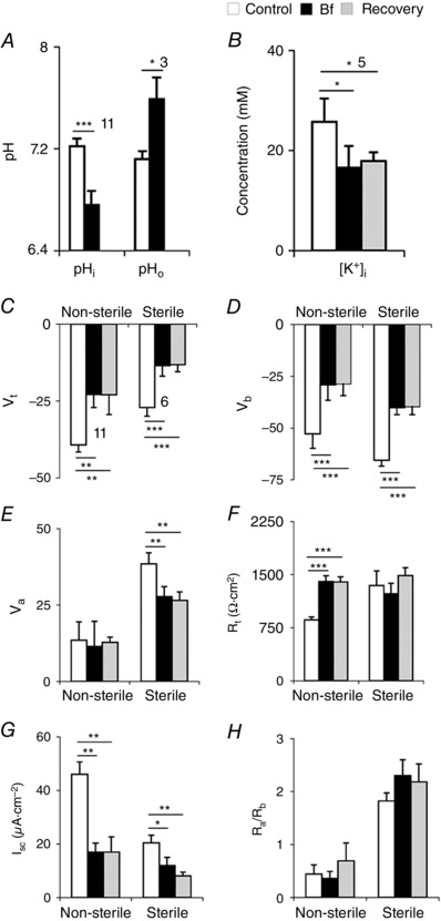
Figure 9. Sterile midguts have highly resistive apical membranes (pooled data).

A–B, bath application of Bf on posterior midguts of non‐sterile larvae. A, Bf inhibited extrusion of H+, resulting in acidification of cells and pHo alkalinized. B, Bf decreased [K+]i in the enterocytes. C–H, pooled data of all Bf (250 nm Bf in Sch‐R) applications in non‐sterile and sterile larval midguts. Sterile larvae have decreased V t (C), V b (D), V a (E), I sc (F) and increased R t (G), with fivefold higher R a/R b ratio (H). Number of experiments (n) is indicated in A and B and n shown in C also applies to D–H. Graphs show mean ± SEM. * P < 0.05, ** P < 0.01, *** P < 0.001.
