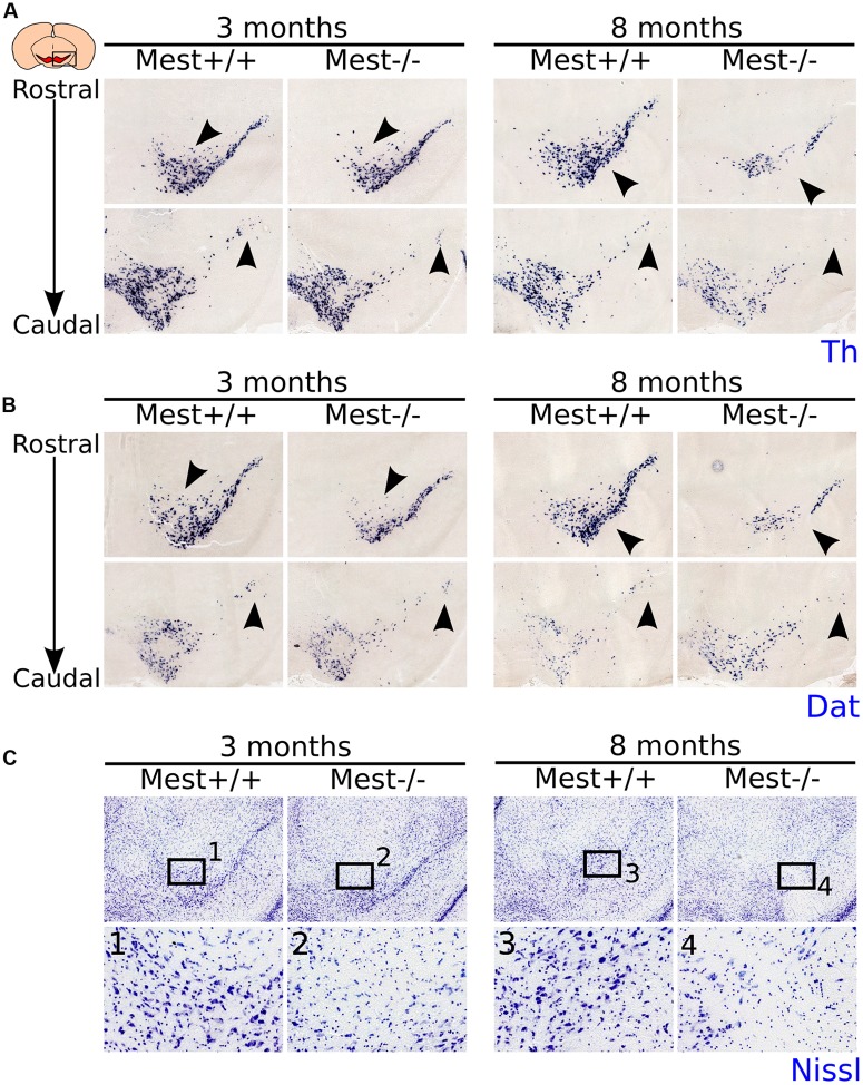
FIGURE 2.
MdDA neurons are lost in the midbrain of adult Mest KO animals. (A) Th-expression (blue) is affected in the SNc of 3- and 8-months old adult KO mice. The loss of Th expression seems to be progressive as more Th+ cells are lost in the 8-months old midbrain (black arrowheads). (B) Dat-expression (blue) is similarly affected in the SNc of 3- and 8-months old adult KO mice as Th-expression (adjacent sections) (black arrowheads). (C) Nissl-staining on adjacent sections at 3- and 8-months old Mest KO and WT controls shows that the loss of Th and Dat is accompanied by a decreased DA cell density in the SNc (black arrowheads), indicating that Mest depletion results in a mdDA cell loss and not in a specific loss of mdDA markers.

