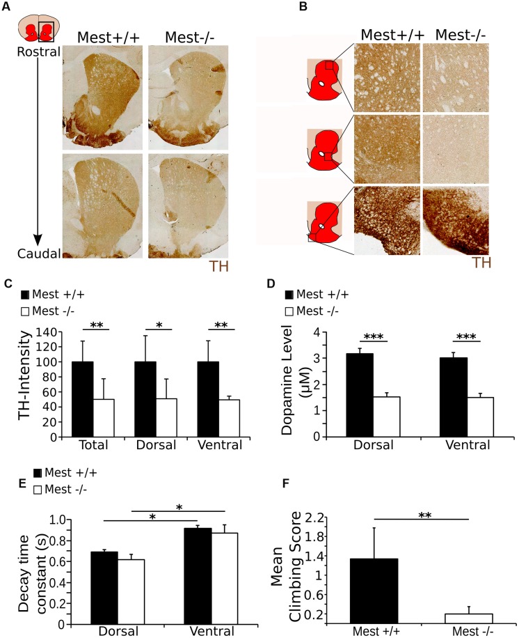
FIGURE 4.
The Mest KO has a 50% lower level of TH in the striatum than the WT control, reflected in the amount of DA release and climbing activity. (A) TH protein (brown) in the striatum of the Mest KO compared to the WT control. (B) TH protein (brown) appears to be decreased throughout the striatum, indicating a general reduction of TH. However, TH presence in the tubercle of the striatum is not apparently altered. (C) Intensity measurements of TH protein level in the striatum of the Mest KO compared to the Mest WT show that the level of TH is significantly reduced in the total striatum with ∼50% (WT n = 4 black bars; KO n = 3 white bars; ∗∗p < 0.01), which is not specific for the dorsal (∼50%; ∗p < 0.05) or the ventral (∼49%; ∗∗p < 0.01) regions of the striatum. WT was set at 100%. (D) Cyclic voltammetry measurements in the striatum of the Mest KO (n = 4 white bars) and WT (n = 4 black bars) show that upon stimulation (500 μA) DA release is reduced with ∼50% in the Mest KO compared to WT controls (∗∗∗p < 0.001). WT animals have a DA release of ∼3 μM in both the dorsal and the ventral striatum, whereas Mest KO animals have a DA release of only ∼1.5 μM in both areas. (E) Time constant of the decay of the DA release in the dorsal and ventral striatum from Mest KO mice (n = 4 white bars) and WT controls (n = 4 black bars) (∗p > 0.05). Decay of DA release is not significantly altered between the WT and KO animals. Also, the characteristics of DA decay of the dorsal and ventral striatum are similar between the WT and KO. (F) Climbing test score. Mest KO (n = 4 white bar) adults show less (WT average score 1.3 compared to KO average score 0.2) activity in the climbing test than WT controls (n = 5 black bar) (∗∗p < 0.01).

