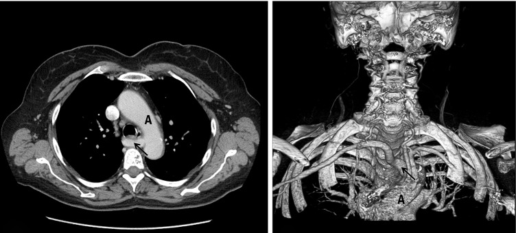
Figure 6.
Computed tomography: (Left) bidimensional scan. The origin of the subclavian artery (arrow) is on the left part of the aortic arch (A) and crosses the posterior wall of the esophagus (transversal section). (Right) Three-dimensional reconstruction. Reproduced with permission from (19).

