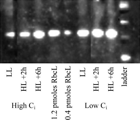
Figure 6.
Representative chemiluminescent blot showing the content of RbcL detected by specific global anti-RbcL antibody in high- and low-Ci before and after the HL shift. At least two lanes per gel were loaded with a Rubisco quantitation standard (AgriSera). Quantitation of RbcL from similar replicate blots is presented in Table I.

