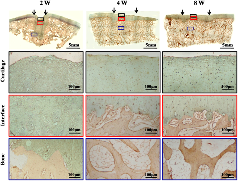
Figure 7. OCN immunohistochemical staining of repaired regions.
In the 2-week group, the entire repaired region presented fibrous tissue with negative staining for OCN. In the 4- and 8-week groups, the repaired regions showed OCN-negative cartilaginous regions (black boxes) and OCN-positive subchondral bone regions (blue boxes) with distinct interfaces (red boxes), similar to the surrounding native structures. Black arrows indicate repaired regions.

