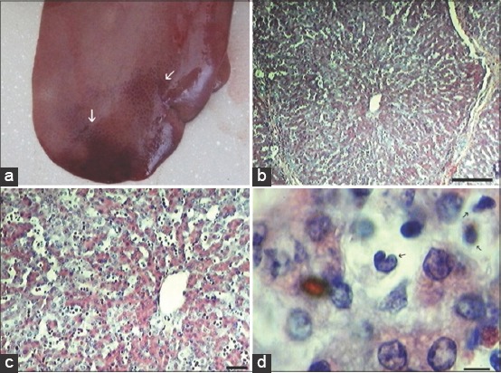
Figure-3.

Liver histopathology: Subcapsular liver hemorrhages (white arrows) from 3 days post-infection (DPI) (a). Erythrocyte accumulation in the areas of liver parenchyma, at 6 DPI (b), scale bar is 250 µm, and (c) Scale bar is 100 µm. At late stage of African swine fever arise infiltrates of leukocytes in portal spaces and sinusoids at 6 DPI (d) arrows show massive leukocytic infiltrates (Scale bar is 10 µm).
