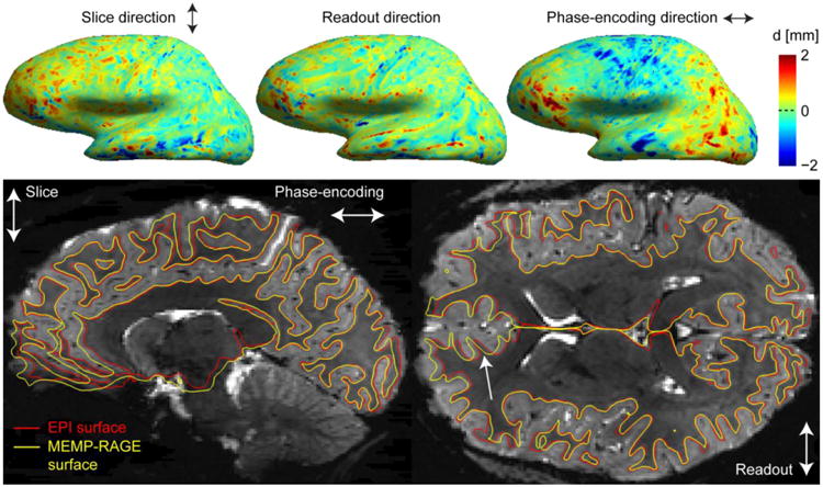
Fig. 12.

Correspondence between MEMP-RAGE and MI-EPI surfaces. Top: The discrepancies of the pial surface placements of MEMP-RAGE and MI-EPI surface reconstructions are shown for the lateral view of the left hemisphere of a representative subject. A vertex correspondence was computed for the surface models, as described in the text, and the discrepancy is shown for the three orthogonal, differently encoded directions. Bottom: Boundary-based affine registrations were computed between the T2* (S0) image and two T1w images, and the surface reconstructions were inverse-transformed to the T2* EPI space; the white surface contours are overlaid on the sagittal and axial slices as indicated. The white arrow points to a location where the MEMP-RAGE surface is misplaced with respect to the T2* EPI slice.
