
Fig. 5.
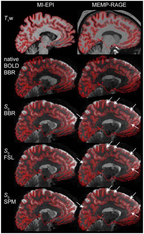
Correspondence between BOLD and S0 images, and performance comparison between different registration methods. The conventional, native BOLD and the derived S0 images registered to the MI-EPI-based T1w and MEMP-RAGE FreeSurfer reconstructions are shown with the corresponding white and pial surfaces indicated with black lines. BBR (boundary-based registration), FSL, and SPM indicate the spatial registration methods and software packages that were used in aligning the T2* data to the T1w anatomical reference images. White arrows point to locations where the boundary-based co-registration slightly outperforms one or both of the volumetric methods.