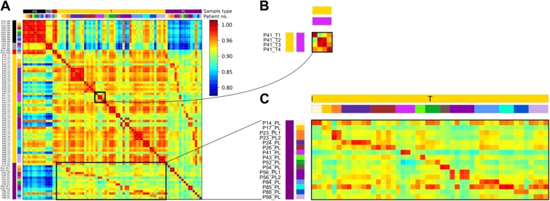
Fig. 2.
DNA methylation of metastasis and primary site from the same patient is highly similar. a Between-sample correlation plot. Sample names are shown to the left of the plot. At the top and the left of the plot are colored sidebars showing sample type and patient identifier. The sidebar to the right of the plot shows the correlation coefficient color key, red being high correlation and blue low correlation. P patient, AN adjacent normal, T primary tumor focus, NL tumor-negative lymph node, PL tumor-positive lymph node. b Enlargement of correlation amongst primary tumor foci in patient 41. c Enlargement of correlation between all primary tumor foci and all positive lymph nodes

