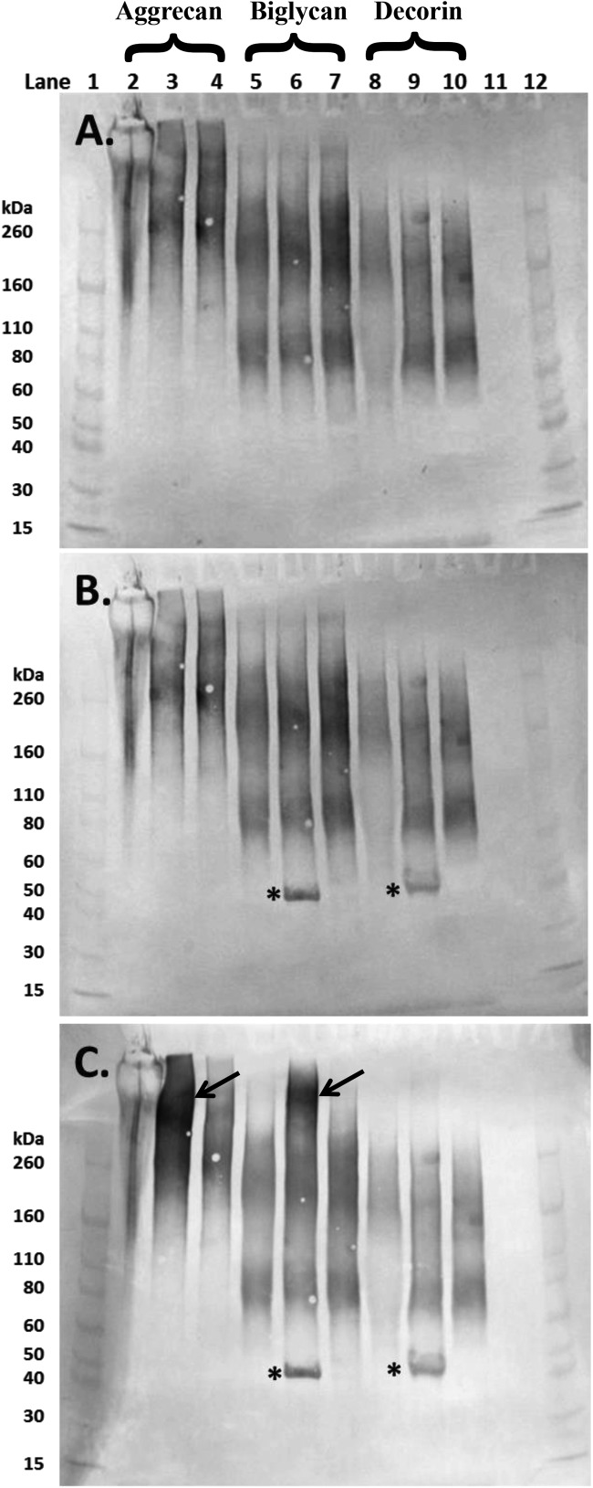
Figure 3. Western blot images of aliquots of purified aggrecan isolated from a human intervertebral disc (lanes 2–4), and the commercially sourced ‘biglycan’ (lanes 5–7) and ‘decorin’ (lanes 8–10) following separation on 4–12% Bis-Tris gel.
PGs in lanes 2, 5 and 8 were undigested whereas those in lanes 3, 6 and 9 and 4, 7 and 10 had been digested with chondroitinase ABC and chondroitinase AC respectively (both 250 mU/ml). Membranes were probed sequentially with monoclonal antibodies to (A) over-sulfated KS heptasaccharides (5D4); (B) 4-sulfated unsaturated disaccharide CS neo-epitopes (2B6) and (C) 6-sulfated unsaturated disaccharide CS neo-epitopes (3B3). Lanes 1 and 12 contain molecular weight standards. The expected molecular weight of the intact core protein of both biglycan and decorin is approximately 45 kDa (*). Aggrecan bands were particularly prominent in lane 3 (4C) following chondrointase ABC treatment (arrow), similar bands were seen in the ‘biglycan’ preparation (lane 6, arrow).

