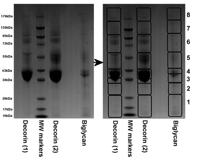
Figure 4. Image of one of the 12.5% SDS-PAGE stained with Coomassie Blue following electrophoretic separation of the commercially sourced ‘decorin’ (1st batches (1) and 2nd batch (2)) and ‘biglycan’ after enzymatic digestion with chondroitinase ABC, keratanase and keratanase II (removing all CS, DS and KS GAG chains).
Protein slices as outlined (1–8) were dissected from each lane and then prepared for MS analyses.

