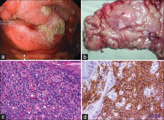
Figure 1.

Diffuse large cell lymphoma stomach. (a) Upper gastrointestinal endoscopy image showing a gastric tumor with central ulceration. The tumor is bulging with increased vascularity. (b) Gross examination of the gastrectomy specimen revealed an ulceroproliferative tumor with central ulceration in the antrum. (c) Photomicrograph of the tumor. Monotonous population of lymphoid cells with moderate to abundant cytoplasm. The cells have hyperchromatic nuclei (H and E, ×400). (d) CD 20 diffuse membrane staining of lymphoid cells (IHC, ×400)
