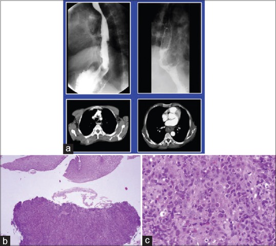
Figure 3.

Diffuse large cell lymphoma esophagus. (a) Barium enema showing narrowing of upper esophagus. Lower window shows computed tomography scan of the same tumor. (b and c); biopsy from the growth reveals esophageal mucosa with a monotonous lymphoid tumor in the submucosal area morphologically consistent with diffuse large cell lymphoma (H and E, ×100, ×400)
