Figure 2.

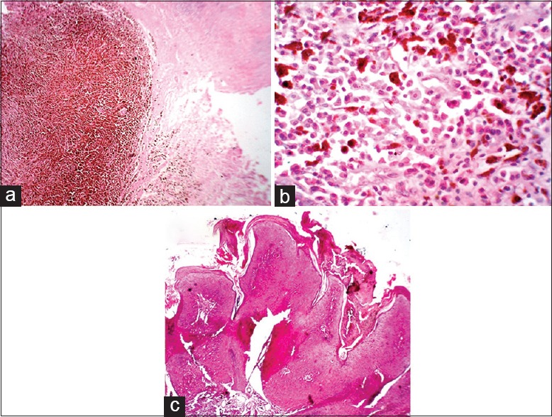
(a) A well circumscribed nodular lesion with excess melanin pigment deposition and irregular arrangement of neoplastic cells (H and E, ×4). (b) Pleomorphic tumor cells with vesicular nuclei with prominent nucleoli seen. Moderate amount of cytoplasm seen. Abundant melanin pigment seen (H and E, ×40). (c) Hypertrophic actinic keratosis: Hyperkeratosis and papillomatosis with prominent cytologic atypia with a moderate lymphocytic infiltrate in the underlying dermis (H and E, ×4)
