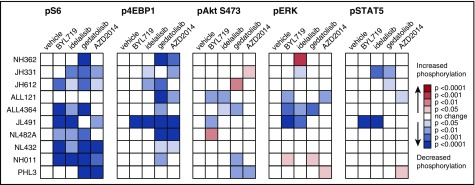
Figure 3.
PI3K pathway inhibitors inhibit signaling phosphoproteins and induce minimal compensatory signaling upregulation. Ph-like ALL PDX models were treated with vehicle, BYL719, idelalisib, gedatolisib, or AZD2014 (n = 3-4 mice per treatment) for 72 hours, then sacrificed at 1 hour after final dose for pharmacodynamic measurement of in vivo target inhibition via phosphoflow cytometry. Heatmap data depict significant changes in phosphoprotein levels in inhibitor- vs vehicle-treated controls using the percentage of FMO+ cells (described in Figure 2) for each PDX model. Colorimetric scale depicts normalization of each model to vehicle controls (white squares; leftmost columns) with statistically significant decreased phosphorylation (blue colors) or increased phosphorylation (green colors) with inhibitor treatment as calculated by ANOVA with the Dunnett posttest for multiple comparisons. FMO+ data for all individual mice are displayed in greater detail in supplemental Figure 5.

