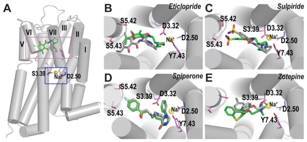
Fig. 2.
Ligand binding mode of Na+-sensitive and -insensitive ligands. (A) The relative locations of the Na+-binding site (blue rectangle) and the ligand binding site (violet rectangle) are shown in the D3R-eticlopride structure.1 The Na+ modeled into the structure is coordinated by the side chain oxygen atoms of Asp(2.50) and Ser(3.39), shown in blue sticks. The Na+ is shown as yellow spheres. The ligand binding modes of the Na+-sensitive ligands, eticlopride (B), sulpiride (C) and zotepine (E), differ in the Na+-bound (green) vs. Na+-unbound (gray) conditions, while those of Na+-insensitive ligand, spiperone (D), are similar in both conditions.

