Figure 2.
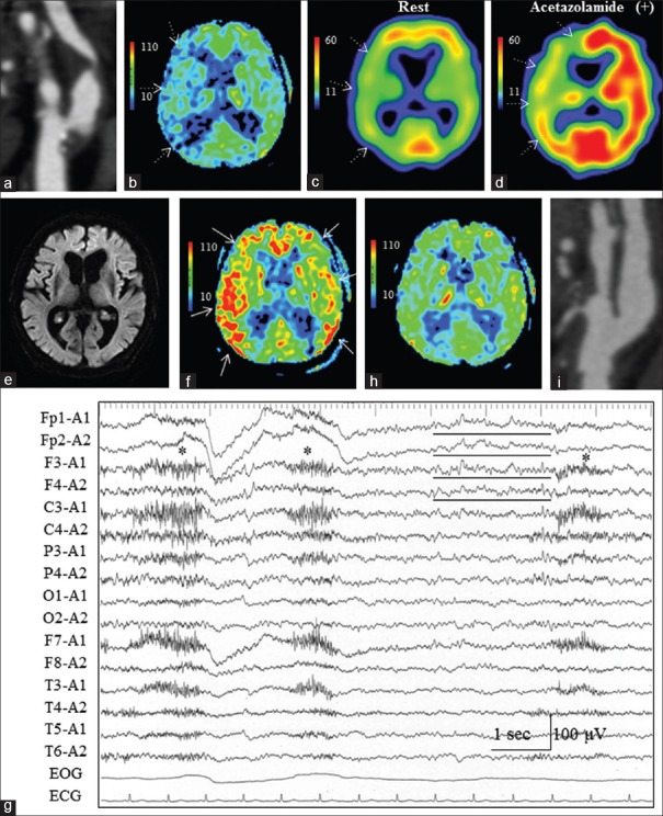
Case 5 (Group A). (a) Preoperative three-dimensional computed tomographic angiography (3D-CTA) revealed severe stenosis of the right internal carotid artery (ICA) at the bifurcation of the common carotid artery. (b) Preoperative magnetic resonance perfusion image with arterial spin labeling (ASL) showed decreased signals in the right middle cerebral artery (MCA) territory (white dotted arrows). (c) Single-photon emission computed tomography with N-isopropyl-[123I] b-iodoamphetamine at rest demonstrated mild reduction of cerebral blood flow in the right MCA territory (white dotted arrows). (d) With acetazolamide loading, impairment of cerebrovascular reserve in the right anterior cerebral artery (ACA) and MCA territories was noted (white dotted arrows). (e) On POD1, diffusion-weighted imaging failed to reveal any de novo ischemic lesions. (f) ASL on POD1 clearly showed increased signals in the bilateral ACA and MCA territories, especially on the right side (white arrows). (g) Electroencephalography on POD1 showed slow-wave activities in the bilateral frontal regions (Fp1, Fp2, F3, and F4 of International EEG 10-20 system, black lines) with poorly organized background activities. Asterisks indicate motion artefact due to restless confusion. (h) ASL on POD14 showed disappearance of the increased signals. The preoperative decreased ASL signals in the right MCA territory were also improved. (i) Postoperative 3D-CTA confirmed that the ICA stenosis was improved

