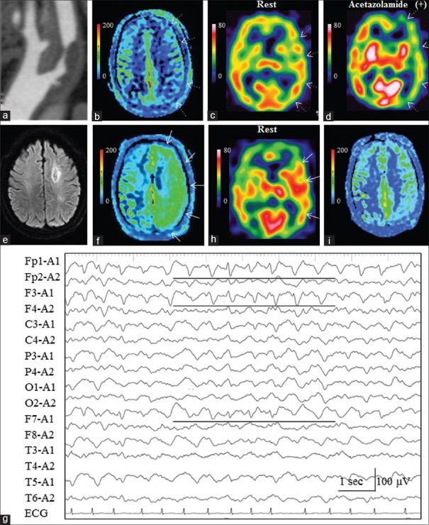
Figure 3.
Case 23 (Group B). (a) Three-dimensional computed tomographic angiography revealed severe stenosis of the left internal carotid artery. (b) Preoperative arterial spin labeling (ASL) showed decreased signals in the left middle cerebral artery (MCA) territory. (c) Preoperative single-photon emission computed tomography (SPECT) image with 99mTc-ethylcysteinate dimer (ECD) demonstrated reduction of cerebral blood flow in the left MCA territory. (d) Acetazolamide challenge depicted impairment of cerebrovascular reactivity in the left MCA territory. (e) On POD1, diffusion-weighted imaging failed to reveal de novo ischemic events, although an old infarction in the white matter of the left frontal lobe was observed. (f) ASL clearly demonstrated increased signals in the operated left hemisphere. A perfusion defect of the old infarction lesion in the white matter of the left frontal lobe was prominent because the ASL signal in the left hemisphere was increased. (g) Electroencephalography on POD1 showed atypical triphasic waves in the left frontotemporal region (Fp1, F3, and F7, black lines) on diffuse slow-wave activities. (h) ECD-SPECT on POD2 still demonstrated hyperperfusion in the left MCA territory. (i) ASL on POD8 showed no laterality in the ASL signals

