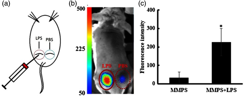
Fig. 8.
Quantification of MMP activity in vivo using the portable imager. (a) An illustration of a skin infection model with subcutaneous injection of LPS and PBS (control). (b) Representative fluorescent image displaying different extents of fluorescent signals emitted by MMP-sensitive fluorescent probe injected subcutaneously with or without the presence of LPS. (c) Quantification of fluorescent intensities demonstrated that LPS treatment significantly increased MMP activity in vivo.

