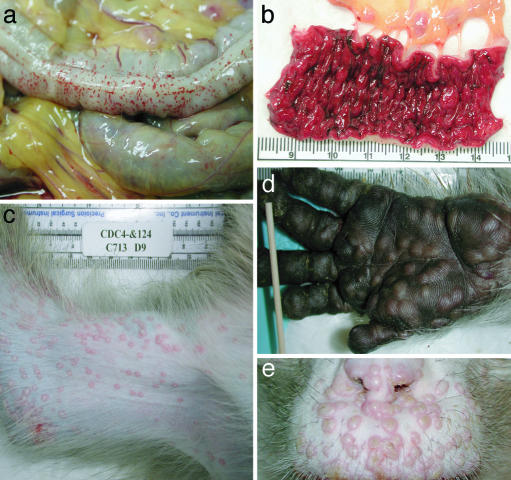
Fig. 2.
Gross lesions associated with smallpox virus infection of monkeys. (a) Hemorrhage on the serosal surface of the distal colon, monkey I-7, 6 days after exposure. Note the diffuse petechial hemorrhages and hemorrhagic, colic lymph nodes. (b) Mucosal surface of the distal colon described in a. Note the severe congestion, hemorrhage, and hemorrhagic colic lymph nodes. (c) Medial surface of the right arm of monkey I-4, 9 days after exposure. Smallpox pustules are predominantly discrete with occasional coalescence. Pock lesions developed synchronously and were more numerous distally, which is consistent with centrifugal distribution. (d). Palmar surface of the left hand of a monkey 11 days after exposure. Discrete and coalescing pustules retained integrity due to heavily keratinized palmar and digital epidermis. (e) Upper lip and nostrils from a cynomolgus monkey 11 days after exposure. Note synchronous development of pustules, some of which have umbilicated. Pustules that formed on the lightly keratinized mucosal surfaces of the nostrils have already ulcerated, resulting in dried exudates.

