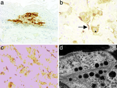
Fig. 3.
Microscopic localization of smallpox virus in tissues of infected monkeys. (a) Immunohistochemical localization of variola antigen in cutaneous epidermis in association with hydropic degeneration and microvesicles, day 3. (b) Variola localization in hepatocytes and Kupffer (arrow) cells of liver, day 6. (c) Variola antigen in association with proximal and distal tubules of kidney, day 6. (d) Electron micrograph of histiocytic cells in spleen, with evidence of replicating virus in association with lamellar membranous bodies and intercellular fibrin.

